In the past, it took a long time to develop a crown as the process includes creating a model of the damaged tooth and the surrounding area, sending it to the dental lab, and waiting for the developed crown to be returned. Patients usually require more than one visit to get a new crown. Implementation of digital imaging technology has changed the situation and now patients can complete the treatment in a single visit.
Digital Imaging Technology for Improved Dental Care
- Digital photography allows patients to see the results of cosmetic dental and restorative dental procedures (how they will look after the procedure). This is accomplished from a series of digital photographs, with the help of digital imaging software and wax models. The process offers huge benefits and provides a high level of communication between the patient and doctor.
- Digital radiographs are much more advanced than films produced from x-ray imaging. In this technology, patients are exposed to a lower degree of radiation. As documents are usually available in digital format, the patient’s digital x-rays or other dental information can be easily stored and archived in a digital file, which can be easily accessed and shared.
- Cone-beam computed tomography is a new application in digital imaging, which is widely used by dental professionals. Data captured from the inner or outer areas of the tooth is used to reconstruct a 3-D image, unlike x-rays that can form only 2-D images. Images (either 2-D or 3-D) are used for dental implant planning, visualization of abnormal teeth, evaluation of the jaws and face, cleft palate assessment, endodontic (root canal) diagnosis, and diagnosis of dental caries (cavities).
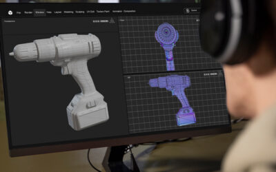
- In computer aided design technology a digital scan is taken of the mouth using a special oral camera. The digital photos of the teeth in 3-D format can be displayed on the computer for the patients to view. Then with the aid of computer software, a crown is designed exactly in the shape and size of the damaged tooth. The digital image is sent to the machine that creates the ceramic crown within two hours.
Digital image capturing and such advanced systems have made digital integration of clinical care, diagnostics and patient communication smooth and efficient. Moreover, special lasers are also being used for detecting cavities and placing filings. It eliminates the need for scalpels, drills and sutures and reduces the patient’s pain and trauma to a great extent.


